Vh endodontic

Vh endodontic
Logo

segunda-feira, 15 de novembro de 2010

Endodontic treatment tooth with pulp nodule

Pulp nodules are alterations of the pulp tissue, presenting as calcified masses, present in sound teeth, decayed, and even traumatized teeth, usually constituting a radiographic finding. Size and varied ways, come to fully block the pulp chamber, hindering access to root canals. (IN: pulp nodules: clinical and morphological)

Case Study:

History: Male, 28 years. Came to the clinic reporting spontaneous pain and throbbing, localized, with peaks during the night and the touch of cold food.

Radiographically: I watched a radiopaque image, indicative of the prosthetic restoration element 46, close to the pulp chamber, with no visible change in the periapical region. It was initially identified an anatomical change in pulp chamber.

Diagnostic hypothesis: irreversible pulpitis.

Proposed treatment: endodontic treatment in one session.

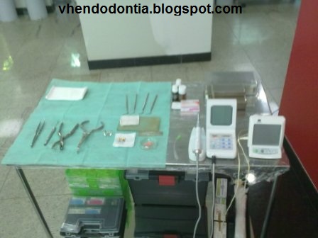
Use of ultrasound Sonic Jet (Gnatus) ET20 with insert. For removal of the lymph pulp, together with the use of an explorer straight (ODUS)
Use of apex locator, Root ZX II (J Morita).
Instrumentation: Motor X-Mart (Dentsply), system files Easy Pro Design.
Final manual instrumentation: Biomechanical file 40 Distal 35 Mesial.
Hypochlorite solution with 5.25%.

Obturation Technique: Hybrid Tagger Endofill endodontic cement (Dentsply).

Infection control

  Set of procedures for supporting endodontic treatment for the maintenance of the aseptic chain, which underpin the philosophy of group therapy aimed at proservation biological integrity of the periapical tissue.
The oral cavity is an area of the body with more diverse microbiota, and 1 ml of saliva from a normal individual in good health, contains about 750 million organisms. Some species of bacteria produce nearly one hundred generations in a period of 24 hours. There are cases where the dental pulp is free of microorganisms and there are times when it is heavily contaminated by them.
The professional should always be aware and meditate on the aphorism.  The provider is not responsible for what is in their field, but for what it brings on.
 So, throughout the endodontic treatment should be made to prevent or minimize the presence of microorganisms in field work during the surgery. (IN: FORP - BASICS OF TREATMENT ENDODONTIC) 
One way of maintaining consegiur this is the organization of the clinic desk and side table.

Using sterile TNT:
 Auxiliary table: sterile instruments and equipment disinfected.

Material disinfected with gauze soaked in alcohol 70%, by friction.

Instrumental sterile

  Table clinic: Instrumental sterile.

Apexification with MTA

Endodontic treatment of teeth with incomplete root formation is complex and requires great professional skill due to occur frequently in young patients. Reduce the number of sessions required for treatment without harming the quality would decrease the damage to the tooth crown due to subsequent reinterventions necessary for the exchange of intra-canal medication, and allow completion of the final restoration soon.
In the treatment of teeth with incomplete root formation and pulp necrosis MTA (Mineral Trioxide Aggregate) can be recommended for making an apical cap (plug), allowing the completion of root canal filling. The mineral trioxide aggregate (MTA) is a cement that was introduced to endodontics by Lee et al. 14 (1993). 14 (1993). Its excellent sealing ability, resistance to microleakage and it can be used in humid environment or with the presence of blood, makes it suitable for use as apical barrier in teeth with incompletely formed apex. Shabahang and Torabinejad (2000) using the MTA as apical barrier, have demonstrated the induction in the formation of hard tissue.
The MTA in conjunction with calcium hydroxide is a valid option for the apexification, bringing main advantage of the speed with which treatment can be completed.  (ANALYSIS IN VITRO VIABILITY OF USING THE MTA AND CALCIUM HYDROXIDE AS PLUG IN TEETH WITH APICAL incomplete root formation. In ABO-GO).


Case Study:


History: Patient special nine years, with cognitive and motor disabilities. Arrived in the clinic with a history of trauma resulting from a fall from their own height, where there was subluxation in the maxilar central incisors.  He was treated at a clinic and made radiographs, where we observed the coronal fracture of the items cited, but without any change in the periapical tissues and bone structures.  After 9 months the patient's mother noted the steady growth and a monthly ball with pus in the region of the element (second report).


Radiographically: Studies in the periapical radiograph, the coronal fractures of the elements 11:21, and noticed a radiolucency in the root element 21.  It was also observed that both elements had to incomplete root formation.
Diagnosis: chronic apical periodontitis.
Proposed treatment: necropulpectomy with apexification with MTA, a single session under intravenous sedation.

Instrumentation final 80.
Hypochlorite solution with 5.25%.
Final irrigation with saline.
Placement of 1 mm calcium hydroxide in the apical region.
Plug 4 mm of MTA.

Anatomic variation - Mandibular Pre Molar - Case report

The anatomy of the root canal system dictates the parameters under which the endodontic treatment is carried out and affects the chances of success. This anatomy of each tooth presents common features and variations very complex.
A radiograph of the tooth can reveal much of the internal anatomy, which combined theoretical knowledge, dictate the size of the drill to be used in surgical access, its direction, the size of the first instrument to be used in root canal and also , which modifications that should be employed to perform endodontic cavity preparation in order to facilitate the location of root canals.
Thus, knowledge of the anatomy of root canals greatly help the professional, since the surgical access to the filling of the channel and is a safe route to achieve much success and avoid unpleasant situations. "(SHORT REVIEW OF HUMAN ANATOMY OF THE TEETH. In Endodontics)

Clinical case:
Histórico : Homem, 23 anos. History: Male, 23 years. Chegou ao consultório relatando dor espontânea e pulsátil, localizada, com maior incidência durante a noite e com contato de alimentos frios ou quentes. Came to the clinic reporting spontaneous pain and throbbing, localized, with peaks during the night and connect with hot or cold food. Dor melhorava, mas não cessava com o uso de analgésico. Pain improved, but did not cease with the use of analgesics.

Radiographically: I watched a radiolucency in the distal element 45, close to the pulp chamber, with no visible change in the periapical region. Foi inicialmente identificada uma variaçao anatômica no formato da raiz do elemento. Was first identified a variation in the anatomical shape of the root element.
Diagnostic hypothesis: irreversible pulpitis.
Proposed treatment: endodontic treatment in one session.

 Use of apex locator, Root ZX II (J Morita).
Instrumentation: Motor X-Mart (Dentsply), system files Easy Pro Design.
Final manual instrumentation: Biomechanical file 35.

 Hypochlorite solution with 5.25%.
Obturation Technique: Hybrid Tagger Endofill endodontic cement (Dentsply).