
One of the most relevant discussions in root canal treatment is what is the distance between the tip and the apex of the dental instrument. Regarding the extent of root canals, precise operation and tooth form the foundation of all subsequent surgical stages of endodontic treatment.
Although several points in this phase of the dental crown can be used as a reference occlusal, the same can not be said of the apical reference, which must be unique and accurate, the foramen. This is the main determinant of referential anatomical apical cleaning, shaping and filling of root canals, which maintains a strong correlation with clinical success, radiographic and histological study of root canal treatment.
Studies show that the apical third has the highest anatomical complications, like buckles and atresia, stressing also the primary variable position of the foramen in relation to the apex. In clinical endodontic, radiographic examination periapical offers an inaccurate position of the endodontic instrument in relation to the foramen, although the use routinely used, and even then, irreplaceable.
The apical constriction is often described as the point from which to extend the root canal filling. PPonce and Fernandez (2003) evaluated histologically the location of the cemento-dentinal junction and the diameter of the apical foramen of the root canal in maxillary anterior teeth. The results showed that the cementum-dentin junction is simply the point at which two converging tissue inside the root canal, which is susceptible to changes depending on each particular clinical situation and about the different extensions of cement into the canal. The apical constriction and apical foramen are not reliable landmarks for the length of apical filling in the end, and its use to calculate the length shutter, can result in damage to periapical tissues.
OThe maximum shutter could affect the success of endodontic treatment, although the odds are worse when there sobreobturation or subobturation significant. (In: The discrepancy between the conventional method of tooth length with standard reference )
Case Study:
History: Male, 25 years. Now is the Integrated Clinic of the FOR, UFG, for rehabilitation of the element 44.
Radiographically: After preparation, proximity to the pulp tissue.

Diagnostic hypothesis: no pathology, treatment indication for prosthetic
Treatment: Treatment in one session.


In detail the position of the apex, with the file path.

Final manual instrumentation: Biomechanical file 40. Solution hypochlorite 1%.
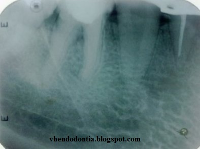
Obturation technique: lateral and vertical condensation

Nenhum comentário:
Postar um comentário