Vh endodontic

Vh endodontic
Logo

segunda-feira, 15 de novembro de 2010

Endodontic treatment of mandibular premolar with 3 degree bend.

The chemical-surgical preparation is aimed at modeling and sanitation, making use of endodontic instruments and auxiliary chemicals instrumentation, working in the root canal system, organic and inorganic structure, providing cleaning and disinfection of the space previously occupied by the pulp, as also continuous conical conformation with higher cervical diameter and smaller apical foramen in keeping the original shape and position, facilitating the realization of the tight seal and three-dimensional (shilder, 1974).
The complexity of the internal anatomy (PINEDA, VERTUCCI, Weiner, HESS, BRAMANTE) and distribution of microorganisms (Shovelton) constitute one of the biggest challenges of Endodontics, especially when related to the curved canals.
NNevertheless, concrescence, crevices and atresias are usually part of the peculiarities of the molars, especially the curved (Schneider).
However, free access to the critical zone of the instrument and the apical canal preparation is a procedure covered with difficulties, which most often result in accidents, among others, excessive wear in the danger zone, with or without perforation (ABOU-RASS ), hourglass-shaped canal, with a smaller diameter in the middle portion of the channel (Buchanan), steps, during apical preparation with or without perforation (Al-Omari; SOUTHARD), besides the possibility of fracture of instruments, due to tensions provided by the bends, as well as diameter, tip design, flexibility of the instrument and instrumentation technique used, variations in the hardness of dentin and obstructions of the channel through the zest of dentin and consequently loss of working length, signal the failure of therapy endodontic.
Many researchers have developed over the last two decades, variations in technical preparation of curved canals from anatomical considerations and their impact described above, with special attention to the technique of cervical ripening (MARSHALL, PAIVA; LOPES).
The cervical ripening may be defined by expanding the diameter at the entrance and the cervical canal, creating a straight access to the middle and apical regions, providing a wear anticurvatura, directed to the areas large or security zones. Consiste numa alternativa para superar a influência da curvatura apical, a partir do desgaste compensatório. It consists of an alternative to overcome the influence of apical curvature, from the wear compensation.
It is the proposition of the authors present a simplified technique for preparing curved canals, using techniques capable of overcoming problems related to the presence of bends and difficulties. (In: SIMPLIFIED TECHNIQUE FOR THE PREPARATION OF CURVED CHANNEL)
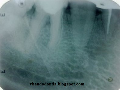

Case Study:
History: Male, 52 years. Came to the clinic with pain, reporting possible fracture of the element 35. On clinical examination revealed a vertical fracture of the crown, bevel, involving the crown of the middle to the vestibular sense lingual vestibule. We observed  the coronal pulp exposed.
Radiographically, was observed on radiographs radiolucent area at the crown and a sharp bend at the root of the element 35 in the region of middle.
Proposed treatment: endodontic treatment in one session.

Rotary instrumentation: Files Easy Pro-designate 20/03. Biomechanics Final: Flexofile 25.
Hypochlorite solution with 5.25%.

Obturation technique: Technique of warm vertical condensation (Schilder 1967).
Endodontic sealer: Endofill (Dentsply)

Radiographic Apex X Anatomy Apex

One of the most relevant discussions in root canal treatment is what is the distance between the tip and the apex of the dental instrument. Regarding the extent of root canals, precise operation and tooth form the foundation of all subsequent surgical stages of endodontic treatment.
Although several points in this phase of the dental crown can be used as a reference occlusal, the same can not be said of the apical reference, which must be unique and accurate, the foramen. This is the main determinant of referential anatomical apical cleaning, shaping and filling of root canals, which maintains a strong correlation with clinical success, radiographic and histological study of root canal treatment.
Studies show that the apical third has the highest anatomical complications, like buckles and atresia, stressing also the primary variable position of the foramen in relation to the apex. In clinical endodontic, radiographic examination periapical offers an inaccurate position of the endodontic instrument in relation to the foramen, although the use routinely used, and even then, irreplaceable.
The apical constriction is often described as the point from which to extend the root canal filling. PPonce and Fernandez (2003) evaluated histologically the location of the cemento-dentinal junction and the diameter of the apical foramen of the root canal in maxillary anterior teeth. The results showed that the cementum-dentin junction is simply the point at which two converging tissue inside the root canal, which is susceptible to changes depending on each particular clinical situation and about the different extensions of cement into the canal. The apical constriction and apical foramen are not reliable landmarks for the length of apical filling in the end, and its use to calculate the length shutter, can result in damage to periapical tissues.
OThe maximum shutter could affect the success of endodontic treatment, although the odds are worse when there sobreobturation or subobturation significant. (In: The discrepancy between the conventional method of tooth length with standard reference )

Case Study:

History: Male, 25 years. Now is the Integrated Clinic of the FOR, UFG, for rehabilitation of the element 44.

Radiographically: After preparation, proximity to the pulp tissue.


Diagnostic hypothesis: no pathology, treatment indication for prosthetic
Treatment: Treatment in one session.
In detail the position of the apex, with the file path.
Final manual instrumentation: Biomechanical file 40. Solution hypochlorite 1%.
Obturation technique: lateral and vertical condensation

Calcified root canal

The conventional endodontic treatment becomes dicícil due to various congenital or acquired. Calcifications can be didactically divided into true and false. The first are the results of action of the odontoblasts, which in some stimulus, they begin to deposit dentin, decreasing or modifying the shape and volume of the pulp cavity.  Among the stimuli we can cite the presence of caries, abrasions and atrições, bruxism and the occurrence of dental trauma. In the latter case, one can verify that the light channel may appear obliterated in the radiographic image is important to note that this calcification is hardly complete, as we have seen, it is the fruit of the action of odontoblasts, there must be at least space occupied by it.  This explains why in some cases, not visualized radiographically the light of the root canal but the tooth has apical lesion. What happens usually is that the lumen of the canal is so narrow that our smallest instrument is too bulky to access it. The call is false calcification resulting from deposition of calcium salts in areas of intra-pulpal microabscesses. True nodules are formed inside the conduit, which may give the impression that this is totally obliterated. Call of false nodules because they are not adhered to dentin walls. These lumps are chronic processes such as periapical pathology, chronic tooth decay and even systemic vascular disorders such as arteriosclerosis. (IN: ENDONLINE)
 
Case Study:
History: Women, 69 years.
 She arrived at the office referred to the treatment endodotico element 24. She reported pain and had no apparent clinical change.  The vitality test positive, exacerbated response. 
Radiographic findings: There was a preparation for fixed prosthesis on vital teeth.


nstrumentation final: file Biomechanics 40, Vestibular, 40, Palatine.
Hypochlorite solution with 5.25%.
Obturation technique: lateral and vertical condensation

Regression of periapical granulomas

The therapeutic principle of any inflammatory disease usually characterized by identifying and eliminating the cause. In the case of periapical granulomas with adequate endodontic treatment characterized by filling the root canal and, if possible, of accessory canals, will be the elimination of the offending agent represented by the microbiota of infected root canals. Bacteria and their products that may remain in small quantities, compared to the potential early aggressive, tend to be neutralized by products used in endodontic therapy, based on biomechanical, along with the irrigating solution, especially antibiotics and calcium hydroxide.
In 60 to 70% of cases of endodontic treatment of teeth with pulp necrosis and chronic periapical success is characterized by regression of the lesion over a few months of the radiographic point of view.However, the ossification process reorganization and apical periodontal tissue begins as soon as adequate endodontic treatment completed. The inflammatory exudate in a few days will be resorbed by inflammatory cells with tissue debris, and microbial cell.  Inflammatory cells will be replaced in a few days and weeks to young proliferating cells arising from surrounding tissues such as periodontal ligament and endosteum. It is expected that over 3 to 4 weeks the tissue is in rapid process of structural reorganization and the inflammatory component is reduced to small pockets in order to clean these areas.Without the mediators of inflammation and its units osteoremodeladoras clasts are demobilized and return to be reabsorbed surfaces covered with cementoblasts proliferating from the surrounding area, returning to form tissue or cementóide osteocemento, reinserting periodontal fibers to restore a normal functional apical periodontium. . However, only a few months after the radiographic area can return to "normal." (IN: DENTAL PRESS) (IN: DENTAL PRESS)

Case Study:

History: Male, 32 years.
He arrived at the office referred to the treatment endodotico element 36. Not refer pain and had no clinically visible changes. The vitality test was negative.

Radiographic findings: There was an extensive carious lesions in the distal crown.
We observed an absence of integrity of lamina dura and a slight radiolucent lesion at the root of it.


Proposed treatment: endodontic treatment in two sessions.
Final manual instrumentation: Biomechanical file 40 Distal 35 Mesial.
Hypochlorite solution with 5.25%.
Systemic medication: Amoxicillin 500 mg for 7 days.

  Obturation Technique: Hybrid Tagger Endofill endodontic cement (Dentsply).

8 months after patient went to a conduit preparation and noted the presence of the hard layer integrates and no periapical changes.

Conventional endodontic retreatment and / or endodontic surgery?

Second Estrela et al. (2001) in determining success in endodontics fulfills criteria, involving clinical examination, radiographic examination and histopathological analysis.  The professional resources available to the longitudinal control, based solely on clinical features (signs and symptoms) and radiographic features.
 Already the second Bender et al, there are some clinical and radiographic criteria representative of endodontic treatment success: 1. No pain and edema,2. Lack of drainage and closure of fistula, 3. Teeth function in normal physiology, 4. Disappearance of periapical bone rarefaction.
According to Friedman and Stabholtz, endodontic point of view, every time there is a failure, the choice rests on two basic tenets: periradicular surgery or conventional endodontic retreatment, which, when properly prescribed, provide a good prognosis.  Since the choice of either option depends on factors such as access to the channel, location and anatomic position of the tooth; involvement with prosthetic pieces, quality of endodontic treatment performed previously, and periodontal involvement.
  Lopes et al. (2004) state that retreatment should be given when initial treatment: Upon radiographic examination, provide inadequate filling; By means of clinical examination, provide exposure of filling a root canal to the oral environment for more than 60 days,  And failure or clinical examination and radiographs.
Even within the third clinical indication: Persistence of symptoms; discomfort to percussion and palpation; fistula or swelling, mobility, inability to chew.  Radiographically, the presence of periapical bone rarefaction in areas previously lacking, including lateral rarefactions; periodontal ligament space increased by more than 2mm; bone repair in the absence of a periradicular rarefaction, an increase of a radiolucent area, no formation of new lamina dura, and evidence of a progression re root resorption.

Case Study:
History: Woman, 27, was referred to our clinic due to the presence of a fistula in the region of tooth 12. Absence of spontaneous pain and also to percussion.
 Radiographically, observed the presence of previous endodontic treatment, with lack of filling material in the apical and lateral condensation disabilities.  Radiolucency suggesting chronic periapical abscess.
17/06/2008 17/06/2008
Proposed treatment: endodontic retreatment  with exchanges monthly calcium hydroxide paste.
17/06/2008 17/06/2008
Removal procedure: with Gates-Glidden drills 4, 3, 2 and 1 through the middle third. Apical hand files, 40, 35 and 30.
 Manual retooling final file 70.
Hypochlorite solution with 5.25%.
Medication Used: calcium hydroxide, iodoform (3:1) and CMCP, saline vehicle.

07/10/2008 07/10/2008
04/11/2008 04/11/2008
Obturation Technique: Lateral condensation with sealer Endofill (Dentsply).
2/12/2008 2/12/2008
2/12/2008 2/12/2008
8/07/2009 8/07/2009

Endodontic retreatment

Endodontics is the science and art that involves the etiology, prevention, diagnosis and treatment of the impact on the periapical region and consequently in the body.  This requires the dentist clinical experience and business sense, and technological resources that provide advantages in cost / benefit ratio for achieving endodontic success, often for the same, being recommended endodontic retreatment before other types of invasive surgeries.
The endodontic retreatment is a procedure performed on a tooth that has undergone a treatment which resulted in a condition that requires a new endodontic treatment to obtain a satisfactory result. The goal of endodontic therapy is to perform a root canal in order to make the treated tooth again functional and comfortable, allowing the complete repair of the support structures.
A condition for the success of endodontic retreatment is proper cleaning of root canals, so should be given to the technique used for removal of filling material, which are usually based endodontic cements with Ca (OH) ² and oxide zinc, folders and gutta-percha, and are sometimes silver cones with prosthetic cement, making it difficult to remove. In the retreatment have to reach the actual length of work and completely remove the filling material, to clean the root canal filling and final.  (In: Articles: http://www.apcd-saocarlos.org.br/arquivos/artigos/desobturacao.pdf )

Case Study:
History: Male, 24 years. Came to the clinic reporting pain induced, localized, with higher incidence during chewing or touch. Região do dente 37. Region of tooth 37.

Radiograficamente: O Radiographically, observed the presence of previous endodontic treatment, with lack of filling material in the canals in mesial and distal canal sobreobturation.  Radiolucency suggestive of periapical lesion.
Proposed treatment: endodontic retreatment.
Use of ultrasound Sonic Jet (Gnatus) ET20 with insert, along with the use of an explorer straight (ODUS).  To remove the core filling resin.
Use of apex locator, Root ZX II (J Morita).
Rotary Instrumentation: Motor X-Mart (Dentsply).
Removal procedure: ProTaper files for removal procedure (D1, D2 and D3).
Roundabout: Files easy pro-designate.
Final manual instrumentation: Biomechanical file 40 Distal 35 Mesial.
Hypochlorite solution with 5.25%.
Obturation Technique: Hybrid Tagger Endofill endodontic cement (Dentsply).


Fourth canal in maxillary first molar

The evolution of endodontics
For years, endodontics has evolved in a way that makes each day that passes, the outcome is becoming increasingly more positive and more favorable prognosis. The numerous techniques, with improved technology and increasingly moving towards the help of endodontic treatment, led to studies that converge in the reasons for endodontic failures.
The use of electronic devices such as apex locator, which brings you greater security in getting the CRD, the NiTi rotary files that help the biomechanical preparation of canals with sharp bends and ultrasound machines that help in the removal of fractured instruments, location channel and the removal of intracanal retainers. However, this does not become an absolute truth, because with the advancement of studies canal treatment was transformed into the treatment of root canals.

This happens because, in a contest X technology channel system, failures are still present in many cases. Several factors are taken into account, however there is one that I consider of great importance. The anatomical variation of the root canal system.

One of the most important anatomical variations and may lead to failure of endodontic treatment is the presence of the fourth canal in maxillary first molar. Most of the first molar mesiobuccal canal presents the palatine (the fourth channel), and this, when untreated, can be the cause of treatment failure.


  According to Ruy Hizatugo, upper molars usually have two root canals in the mesio buccal - MV and MP channel. Histologically, the polls have shown MP channels in approximately 100% of cases. However, clinically, the MP channel is identified and treated in about 80% of cases without using surgical microscope and about 90% of cases with the aid of a microscope and ultrasonic tips.

So why has not failed in first molars that was not handled the mesio palatine canal? This happens because only 14% of cases: two independent channels in the mesiobuccal root.